Joint pain and disease in cystic fibrosis – A practical approach
Introduction
Episodes of joint pain are well recognised in cystic fibrosis (CF), usually starting after ten years of age, and occurring in about five to ten percent of patients (Lawrence et al, 1993).
The commonest form of joint pain in CF is an arthritis that mostly affects the large joints, for example the knee, ankle, wrist, elbow and shoulder. This is sometimes referred to as CF associated arthritis. Episodes usually last less than a week but can be quite disabling. Acute onset at a young age is often sudden with widespread joint pains and general ‘flu-like’ symptoms. Patients often cannot walk because of leg pains and just want to stay in bed. Sometimes the attacks are associated with high swinging fevers and skin rashes.
Most patients rapidly respond to non-steroidal anti-inflammatory drugs (NSAIDs) such as ibuprofen (Brufen®). Short courses of prednisolone may be needed in a minority of patients. Occasionally more aggressive and potentially toxic treatments are necessary. These patients require the expertise of a consultant rheumatology specialist.
The arthritis follows a remitting and relapsing course. Symptoms mostly completely disappear between attacks. X-rays tend to show no abnormalities. Although in most cases there is no permanent damage to the affected joints some have progressed to an erosive arthritis with bone destruction.
A second form of bone and joint disease is called hypertrophic pulmonary osteoarthropathy (HPOA). It is usually found in young adult patients and has an insidious onset. Pain, which is generally mild at the beginning, may increase to a continuous ache. The clinical picture may vary from a minimally swollen joint to tender, warm and swollen joints resembling those seen in rheumatoid arthritis. Symptoms are often worse in cold weather.
Hypertrophic pulmonary osteoarthropathy also occurs in a variety of other lung diseases. In CF it is responsible for finger clubbing, swelling of the ends of the long bones with resulting tenderness, for example those in the forearm, and pain or swelling of large joints like the ankle or knee. In most patients the joint symptoms are symmetrical, for example affecting both wrists, both knees or both ankles. X-rays show the periosteum, the tissue covering the bone, elevated from the bone surface and some new bone formation. These x-ray changes may not appear for several months after the clinical signs and pain. The changes can slowly progress and can result in permanent bony destruction.
As with classic CF associated arthritis, symptoms usually respond to non-steroidal anti-inflammatory drugs with considerable relief of pain and swelling. Again corticosteroids may be needed for cases resistant to these treatments. Rest of the affected limb and local heat treatment can help reduce pain, stiffness and swelling. HPOA may also resolve if the underlying chest disease is intensively treated.
Both the above forms of arthritis can be associated with acute respiratory exacerbations. This may reflect the upgraded immune response that accompanies acute infections, the arthritis being another manifestation of a hyperactive immune system.
Unlike HPOA, CF associated arthritis is not more common in patients with more severe chest disease. Interestingly clubbing, a sign of HPOA, tends to slowly resolve after lung transplant.
We must remember that patients with CF are susceptible to joint disease unrelated to their underlying illness and that these may need to be excluded by appropriate tests. Many patients with CF are on multiple medications and the possibility of the arthritis being a reaction to one of these drugs, for example ciprofloxacin or cimetidine, should always be considered.
The following approaches should be considered for patients with joint involvement resistant to treatment with NSAIDs:
• Look for symptomatic HPOA, not easy in a population with finger clubbing. HPOA produces pain at the wrists, ankles and knees without clinical synovitis but with occasional effusions. There may be tenderness along the long bones and X-ray or bone scan evidence of periostitis. Elevating the limb may reduce the pain. The treatment (which is often unsatisfactory) is NSAIDs and vigorous treatment of the underlying condition, e.g. a respiratory exacerbation
• Ask again about any family history of joint disease
• Treat as for the most closely resembled classical rheumatological syndrome
• Refer for an expert rheumatology opinion. The patient will likely have further investigations, and treatment with corticosteroid sparing agents such as methotrexate or azathioprine may be considered. Other possible treatments include sulphasalazine and hydroxychloroquine
Taking a history
A physician wishing to treat an individual with cystic fibrosis and joint pain should ask themselves the following questions “Is this pain the result of hypertrophic pulmonary osteoarthropathy (HPOA), an inflammatory condition or a minor mechanical abnormality revealed by muscle weakness?
Early morning stiffness (EMS) of the joints differentiates chronic inflammatory (EMS>45 minutes) from non-inflammatory disease (EMS a few minutes, perhaps 5-10 minutes). In acute arthritis pain predominates over stiffness.
Enquiry should be made for psoriasis (Benjamin & Clague, 1990) and the classical reactive arthritis triggers of infective viral contacts, diarrhoea and sexually acquired infection (Hind, 1982).
The nature and timing of rashes may be revealing. The rash of Still’s disease usually appears in the evening, is associated with pyrexia and is gone by the morning (Larson, 1984). The rash of parvovirus B19 lasts a day or so (Woolf et al, 1989). It may be easier to identify “slapped cheek ” rashes in family contacts than in the patient who may suffers regular rashes.
Drug history
Young people hardly think of acne medications as being ‘drugs’ and may fail to mention that they are on minocycline, which can cause systemic lupus erythematosis (SLE) (Gough et al, 1996). Fluoroquinolone can induce a peritendinous thickening, which can raise suspicions of joint disease but can be differentiated clinically on MRI (Loeuille et al, 1996). Fluoroquinolone induced Achilles tendon rupture may occasionally occur (Ribard et al, 1992). If the calf is compressed with the patient prone and the knee flexed to 90 degrees, there is plantar flexion of the foot; this is lost in rupture of the Achilles tendon.
Family history
Taking a good family history is critical for defining the probability that your patient has a non-CF associated condition.
Gout
There is not a single case of gout in CF reported in the journals covered by “Medline” although we have seen cases in our practice. Uric acid excretion was significantly raised with older pancreatin preparations but this is not a problem with those used at present (Sack et al, 1980; Bohles & Milchalk, 1982; Wiersbitzky et al, 1989). Diagnosing a red joint as gout is therefore unlikely to be correct unless sodium urate crystals are identified in aspirates from the joint.
CF associated arthritis
Cystic fibrosis associated arthritis (CFAA) is described as episodic (lasting less than a week), non-erosive, involving large joints and responding to NSAID (Johnson & Knox, 1994). This is reassuring but patterns of disease can change and if the episodic disease transforms into a continuous small joint arthritis that does not respond to NSAID, it is less likely to be diagnosed as a CF associated arthritis.
Some people with CF suffer from a NSAID resistant synovitis and subsequent metocarpo-phalangeal (MCP) joint space narrowing and subluxation (Rush et al, 1986).
What is the physician to do with the patients who have joint involvement which is resistant to NSAID and a modest increase in steroid dose?
a) Look for symptomatic HPOA, which is not easy in a population most of whom have finger clubbing. HPOA produces episodic pain at the wrists, ankles and knees without clinical synovitis, but with occasional effusions. Exacerbation of pain may precede pulmonary deterioration, and HPOA is more frequent in the later stages of the lung disease. There is tenderness along long bones and X-ray or bone scan evidence of periostitis. Elevating the limb may lessen pain. The treatment (which is frequently unsatisfactory) is NSAID and vigorous treatment of the underlying condition.
b) Re-examine the family history for joint disease.
c) Treat as for the most closely imitated classical rheumatological syndrome.
d) Requires the ability to elicit certain physical signs. These are the same physical signs required to diagnose non-CF associated joint disease such as rheumatoid arthritis, hypermobility and ankylosing spondylitis (Forouzesh & Bluestone, 1979). A screening test for hypermobility in adults is the ability to passively hyperextend the little finger to 90 degrees at the metacarpophalangeal joints (MCP). There is a formal score to pursue if this is positive (Grahame et al, 2000). The modified Schober index screens for limited lumbar movement (Moll & Wright, 1971) which in a young person who has back pain and stiffness that improves with exercise may indicate ankylosing spondylitis. To perform the test, a horizontal line is drawn in the midline, on the patient’s skin at the level of the “dimples of Venus” A mark is made 10 cm above this line and another 5 cm below creating a 15 cm gap between the two most distant marks. The patient attempts to touch his/her toes. As an approximation the distance between the marks should increase by at least 5 cm – anything less indicates limitation of lumbar spine movement and justifies a search for other manifestations of ankylosing spondylitis (Moll & Wright, 1971).
Inflammation of the knee
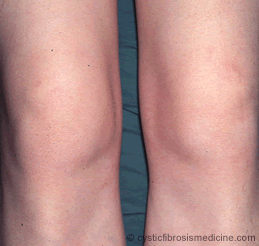
Inflammation of the knee is easily identified as it causes the anterior surface of the patella to be warmer than the mid-tibia. Normally the anterior patella is cooler than the mid-tibia (assuming no heat packs, embrocation, severe varicose veins etc). The sense of diagnostic triumph at finding an “arthritic” effusion may be short lived when it is realised that HPOA as well as arthritis can generate small knee effusions, though these are relatively cool and the synovial fluid contains few white blood cells (Phillips & David, 1986). In patients with CF and acute monoarthritis, sepsis requires exclusion by aspiration, gram staining and culture of the aspirate as in the rest of the population.

Figure 1. Inflammation of the knee with small effusions
Back pain
Sudden onset of severe back pain may represent vertebral collapse in these patients who are predisposed to osteoporosis.
Laboratory Test
Acute phase proteins are difficult to interpret in the presence of chronic lung inflammation.
IgM rheumatoid factor has been reported positive in between 6% and 20% of people with CF; IgG rheumatoid factor was positive in 88%. ANA is usually negative. The patterns of arthritis in IgG sero-positive patients do not necessarily mirror those of IgM sero-positive rheumatoid arthritis (Schiotz et al, 1979; Phillips & David, 1986).
ANCA specific for bactericidal/permeability increasing protein (BPI) is a very early finding in CF. Titres of antibody against proteinase 3 (PR3) are associated with age and pseudomonas infection in a paediatric population. c-ANCA against PR3 or BPI is more common than p-ANCA (Sediva et al, 1998).
It is probably wrong to start a cyclophosphamide/steroid regimen in a patient with CF who has skin vasculitis and ANCA positivity unless severe systemic complications (especially renal or CNS) has been demonstrated. It is worth remembering that the less serious manifestations of even Wegener’s granulomatosis can be controlled by methotrexate with prednisolone (Stone et al, 1999).
Treatment For CF associated arthritis
For CFAA, NSAID should be tried first and if these fail, an increase in corticosteroid dose should be tried. If the doses required is likely to produce adverse effects a steroid sparing agent such as methotrexate or azathioprine will be required. There is very little data on treatment of CFAA that does not respond well to NSAID.
Sulphasalazine is attractive except in cases with a systemic onset similar to Still’s disease. In Still’s disease there is an increased incidence of inefficacy and adverse effects (Jung et al, 2000). A significant positive speckled ANA titre also makes sulphasalazine less attractive as the clinical picture and immunology can be shifted towards a lupus-like syndrome (Gunnarsson et al, 2000).
Hydroxychloroquine is a slow acting agent, again data is lacking but it may be useful when the patient is able to wait four to six months for a response.
Weekly oral treatment with methotrexate (starting at 7.5 mg weekly and increasing to 20 mg by approximately 5 mg increments with a dose of 20 mg folic acid 24 hrs after the methotrexate) can reduce steroid requirements and resolve inflammation. If it is suspected that inefficacy is caused by poor absorption (the author is unaware of studies of methotrexate pharmacokinetics in cystic fibrosis), the intramuscular route can be used (Jundt et al, 1993). The potentially fatal pneumonitis (Van der Veen et al, 1995) and the hepatic fibrosis that it can produce are a concern. There is a convention that initially fortnightly Hb, WBC, platelet count and liver function tests are required. Methotrexate should be stopped if neutrophils fall below two, but ALT may be allowed to increase to three times normal before drug withdrawal. Treatment should be monitored closely by specialists conversant with the drug. Intramuscular therapy in general has advantages in a patient group with variable absorption of oral drugs.
Other systemic therapy such as gold is supported by anecdote rather than formal trial data (Benjamin & Clague, 1990). There are no reports of the use of tumour necrosis factor antagonists (etanercept, infliximab) in cystic fibrosis associated arthritis.
Key points
• Joint pain is a recognised complication of CF
• Arthritis usually affects the large joints
• HPOA is a second form of bone and joint disease seen in CF
• Symptoms usually respond to NSAIDs
• Remember that people with CF may have joint disease that has nothing to do with their CF
• There should be a low threshold for referral to a rheumatology specialist
References
Benjamin CM, Clague RB. Psoriatic or cystic fibrosis arthropathy? Difficulty with diagnosis and management. Br J Rheumatol 1990; 29: 301-302. [PubMed]
Bohles H, Michalk D. Is there a risk of kidney stone formation in cystic fibrosis? Helvetia Paediatrica Acta 1982 ; 37: 267-72. [PubMed]
Forouzesh S, Bluestone R. The clinical spectrum of ankylosing spondylitis. Clin Orthop 1979; 143: 53-58. [PubMed]
Gough A, Chapman S, Wagstaff KA, et al. Minocycline induced autoimmune hepatitis and systemic erythematosus like syndrome. Br Med J 1996; 312: 169-172. [PubMed]
Grahame R, Bird H, Child A, et al. The British Society for Rheumatology Special Interest Group on Heritable Disorders of Connective Tissue criteria for the benign joint hypermobility syndrome. The Revised (Brighton 1988) criteria for the diagnosis of BJHS. J Rheumatol 2000; 27: 1777-1779. [PubMed]
Gunnarsson I, Nordmark B, Hassan Bakri A, et al. Development of lupus -related side-effects in patients with early RA during sulphasalazine treatment -the role of IL-10 and HLA. Rheumatology 2000; 39: 886-893. [PubMed]
Hind CR. Reactive arthritis. Postgrad Med J 1982; 58: 31-37. [PubMed]
Hoiby N, Wiik A. Antibacterial precipitins and autoantibodies in serum of patients with cystic fibrosis. Scand J Resp Dis 1975; 56: 38-46. [PubMed]
Johnson S, Knox AJ. Arthropathy in cystic fibrosis. Respiratory Medicine 1994; 88: 567-570. [PubMed]
Jundt JW, Browne BA, Fiocco GP, et al. A comparison of low dose methotrexate bioavailability, oral solution, oral tablet, subcutaneous and intramuscular dosing. J Rheumatol 1993; 20: 1845-1849. [PubMed]
Jung JH, Jun JB, Yoo DH, et al. High toxicity of sulfasalazine in adult-onset Still’s disease. Clin Exp Rheumatol 2000; 18: 245-248. [PubMed]
Larson EB. Adult Still’s disease. Evolution of a clinical syndrome and daiagnosis, treatment, and follow-up of 17 patients. Medicine 1984; 63: 82-91. [PubMed]
Lawrence JM, Moore TL, Madson KL, et al. Arthropathies of cystic fibrosis: case reports and a review of the literature. J Rheumatol 1993; 20 (suppl 38): 12-15. [PubMed]
Lawson TM, Amos N, Bulgen D, et al. Minocycline-induced lupus clinical features and response to rechallenge. Rheumatology 2001; 40: 329-335. [PubMed]
Loeuille D, Gillet P, Netter P. Fluoroquinolone induced arthralgia and magnetic resonance imaging. J Rheumatol 1996; 23: 1313-1314. [PubMed]
Moll JMH, Wright V. Normal range of spinal mobility. An objective clinical study. Ann Rheum Dis 1971; 30: 381-388. [PubMed]
Phillips BM, David TJ. Pathogenesis and management of arthropathy in cystic fibrosis. J Roy Soc Med 1986; 79: 44-50. [PubMed]
Ribard P, Audisio F, Khan MF, et al. Seven Achilles tendinitis including 3 complicated by rupture during fluoroquinolone therapy. J Rheumatol 1992; 19: 1479-1481. [PubMed]
Rush PJ, Shore A, Coblentz C, et al. The musculoskeletal manifestations of cystic fibrosis. Semin Arth Rheum 1986 ; 15 : 213-225. [PubMed]
Sack J, Blau H, Goldfarb D, et al. Hyperuricosuria in cystic fibrosis in cystic fibrosis patients treated with pancreatic enzyme supplementation. A study of 16 patients in Israel. Isr J Med Sci 1980; 16: 417-419. [PubMed]
Schiotz PO, Egeskjold EM, Hoiby N, et al. Autoantibodies in serum and sputum from patients with cystic fibrosis. Acta Path Micobiol Scand -Section c, Immunology 1979; 87: 319-324. [PubMed]
Sediva A, Baryunkova J, Kolarova I, et al. Antineutrophil cytoplasmic antibodies (ANCA) in children with cystic fibrosis. J Autoimmunity 1998; 11: 185-190. [PubMed]
Stone JH, Tun W, Hellman DB. Treatment of non-life threatening Wegener’s granulomatosis with methotrexate and daily prednisolone as the initial therapy of choice.J Rheumatol 1999; 26: 1134-1139. [PubMed]
Wiersbitzky S, Ballke EH, Wolfe E, et al. Uric acid serum concentrations in CF children after pancreatic enzyme supplementation. Padiatrie Grenzgebiete 1989; 28: 171-173. [PubMed]
Woolf AD, Champion GV, Chishick A, et al. Clinical manifestations of human parvovirus B19 in adults. Arch Int Med 1989; 149: 1153-1156. [PubMed]
van der Veen MJ, Dekker JT, Dinant HJ, et al. Fatal Pulmonary fibrosis complicating low dose methotrexate therapy for rheumatoid arthritis. J Rheumatol, 1995; 22: 1766-1768. [PubMed]
